What is Nuchal Translucency Measurement?

Down Syndrome is the most common chromosomal abnormality. All pregnant women are evaluated in terms of the risk of their babies carrying this syndrome between the 11th and 14th weeks of pregnancy. This test is called a double test. The test has several stages. First, the nuchal translucency is measured with ultrasound, then a special blood test is performed. The pregnant woman’s personal characteristics, the ultrasound measurement and the blood test results are combined in a special computer program to calculate the baby’s Down Syndrome risk.
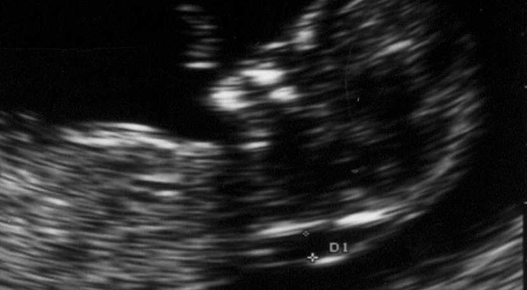
Studies have determined that babies with chromosomal abnormalities, especially those with Down syndrome, have thickening in the subcutaneous area of the neck. This subcutaneous edema is seen much more between 11-14 weeks in the womb. Based on this information, the nuchal translucency of all babies is measured with ultrasonography during this period in the womb. Doctors receive special training in order to perform the measurement. The baby being in the appropriate position and the ultrasound device that performs the procedure having sufficient image quality are necessary conditions. Although the procedure is usually performed by looking at the abdomen, sometimes it may be necessary to perform a vaginal ultrasonography.
When the nuchal translucency measurement value is over 2.5 mm, the risk of Down syndrome increases. However, nuchal translucency alone does not diagnose Down syndrome. Other genetic defects, infections, and heart anomalies can also present with increased nuchal translucency in the womb. If very high values are observed, it is recommended that a procedure called CVS (chorion villus biopsy) be performed to diagnose without delay. This test provides information about the baby’s genetic structure at an early stage. From the 16th week of pregnancy, the baby’s chromosome structure can be revealed by performing an amniocentesis procedure.


